

Recently, Professor Yu Zhangsheng, founder of Intelligent Medicine Original led his team in collaboration with international pharmaceutical companies to publish an article titled "..." online in the Chinese Academy of Sciences' Q1 journal *npj precision oncology*.“Deep learning for oncologic treatment outcomes and endpoints evaluation from CT scans in liver cancer”This research presents the findings of a deep learning-based tumor progression assessment tool that can automatically evaluate tumor burden and new lesions on longitudinal three-dimensional CT images of patients, thereby providing independent, stable, and objective disease progression assessment results for clinical trials in oncology. Xia Yujia and Zhou Jie, doctoral students at the School of Life Sciences and Biotechnology, Shanghai Jiao Tong University, are co-first authors of this paper, while Professor Yu Zhangsheng, Dr. Zhao Shuai from Xinhua Hospital affiliated with Shanghai Jiao Tong University School of Medicine, and Dr. Zhang Jin from PiHealth are co-corresponding authors.
Objective response rate (ORR) and progression-free survival (PFS) are commonly used outcome measures in phase II/III clinical trials of anti-tumor drugs. The accuracy of these measures depends on the accurate assessment of drug treatment results. RECIST v1.1, the standard for evaluating the efficacy of treatment in solid tumors, is currently the guideline used to assess tumor response. It defines a standardized method for assessing efficacy response based on changes in tumor burden. Based on regular imaging follow-ups of patients during treatment, changes in the tumor in follow-up images are compared with baseline images to assess disease remission, stabilization, and progression.
In clinical practice, radiologists typically need to identify lesions, measure tumor burden, and differentiate new lesions in 3D CT imaging sequences to monitor drug treatment response—an extremely time-consuming and labor-intensive task. Furthermore, the subjectivity of image interpretation can lead to discrepancies in image assessment between different radiologists. Studies have shown that the inconsistency rate between two interpreters can reach 23% to 46%. Inaccurate efficacy assessments can lead to flawed medical decisions, affecting treatment outcomes and shortening patient survival. Inaccurate assessments of drug treatment outcomes can result in effective drugs not being marketed or ineffective drugs being marketed. Therefore, improving the standardization and consistency of image assessment is a crucial issue that urgently needs to be addressed.
This research aims to develop an automated tumor progression assessment model based on deep learning. This model transforms the assessment process of clinical radiologists into a deep learning model workflow. It first accurately segments the tumor from 3D CT images, then calculates the overall tumor burden change based on baseline and follow-up images, identifies new lesions, and finally generates an assessment of tumor progression. The model employs a multi-task learning framework, simultaneously optimizing the classification tasks for tumor segmentation and progression assessment, and integrating prior medical knowledge and the correlation between longitudinal images, thereby improving the accuracy of existing liver tumor segmentation models in progression assessment.
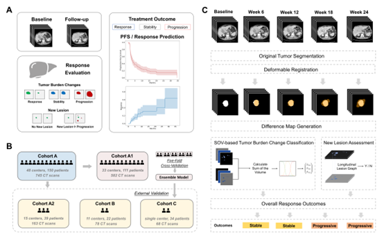
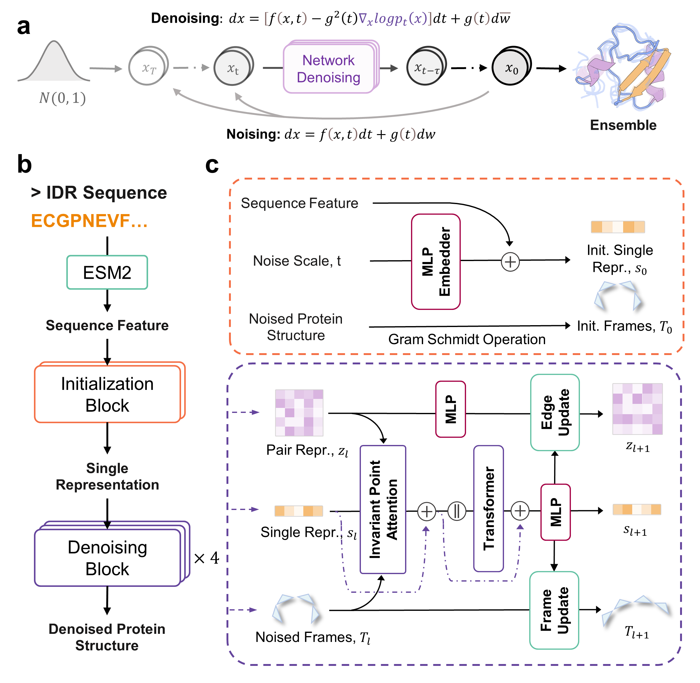
Figure 1. Schematic diagram of this research work
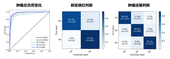
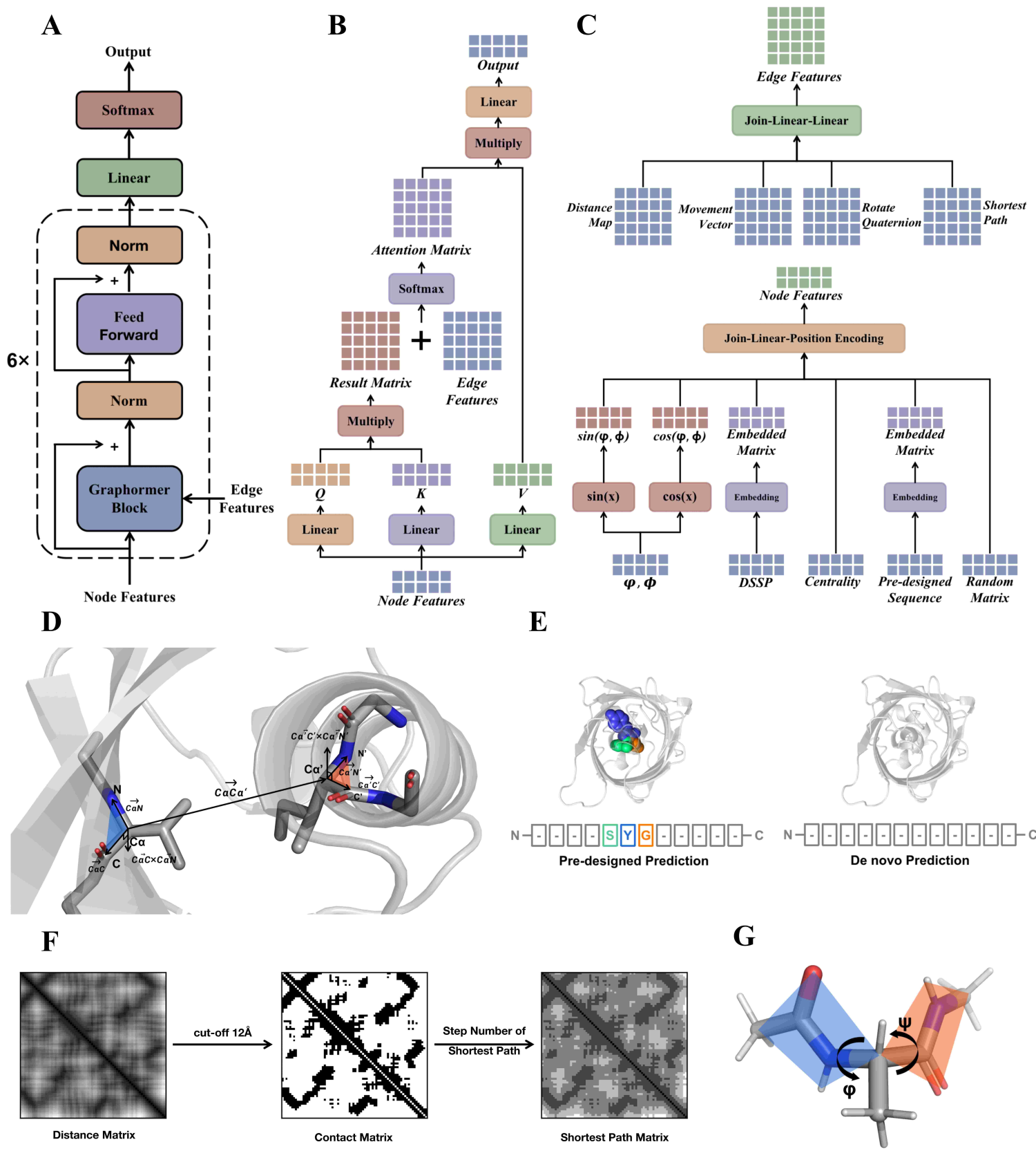
Figure 2. Model accuracy validation results
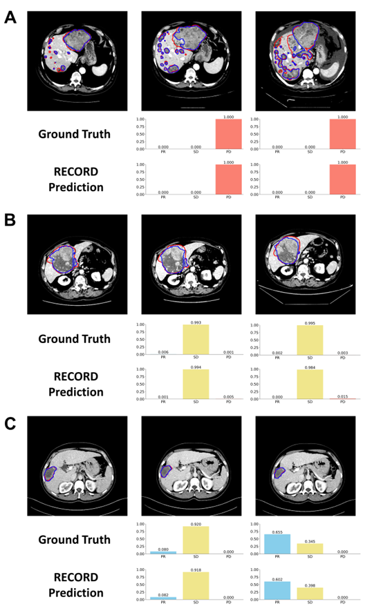
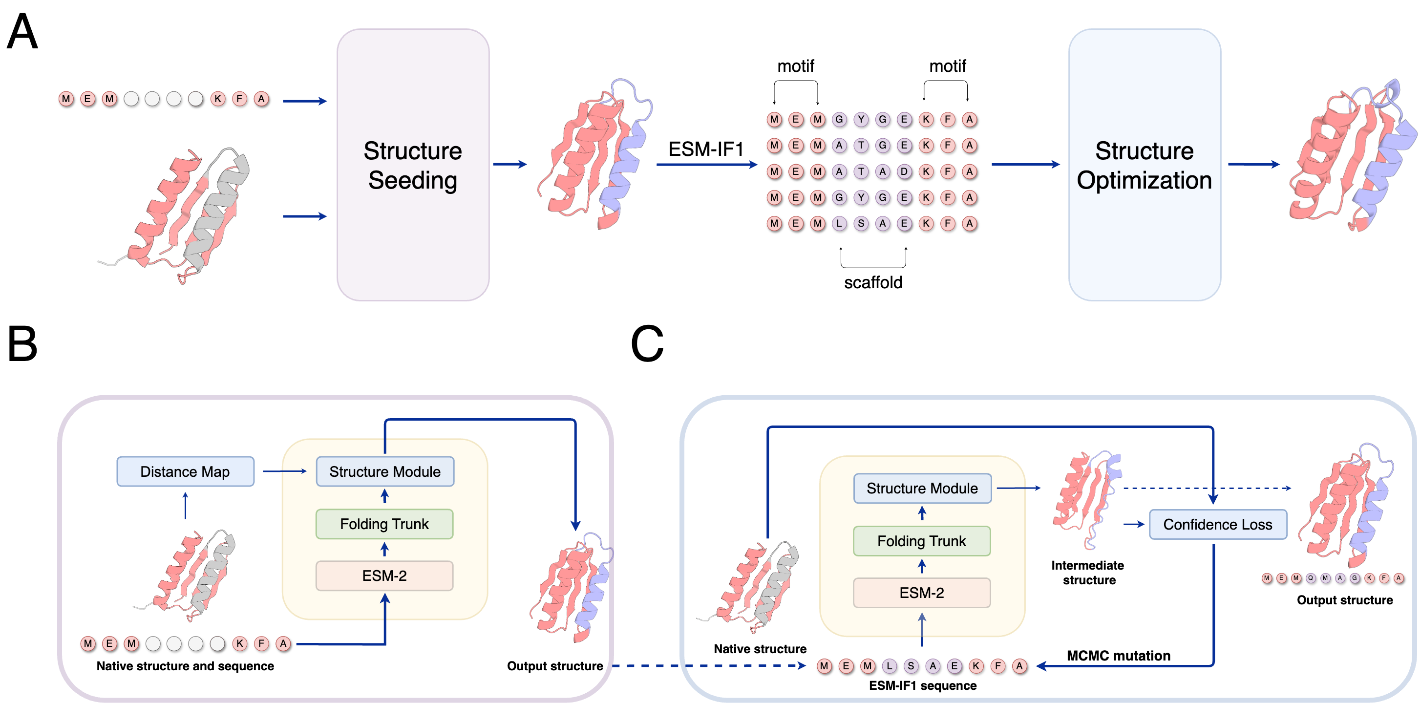
Figure 3. Comparison between model predictions and actual judgments
The constructed model underwent five-fold cross-training in a multinational, multicenter clinical trial cohort. The integrated five-fold model was then evaluated on three heterogeneous independent external validation sets. These sets encompassed primary hepatocellular carcinoma, metastatic hepatocellular carcinoma, and liver cancer under mixed immunotherapy scenarios encountered in international clinical trials and real-world clinical practice. The model's effectiveness was validated in three aspects: First, in baseline-follow-up image analysis, the model achieved accuracies of 0.927, 0.982, and 0.882 in the three external validation cohorts, significantly outperforming existing liver tumor models in progression prediction accuracy. Second, at the patient endpoint outcome level (i.e., progression-free survival and drug response time), the model's predicted outcome time showed a concordance of 0.958 with the actual outcome time. Finally, regarding the correlation with overall survival, the disease progression status assessed by the model showed a stronger correlation with patient overall survival than manually interpreted disease progression results. The developed model can be used for independent image interpretation in clinical trials, thereby reducing the subjectivity of physician assessments and promoting consistency in treatment efficacy assessments across medical centers.
Original link:《Deep learning for oncologic treatment outcomes and endpoints evaluation from CT scans in liver cancer》

 WeChat Customer Service